Get expert, compassionate care for hydrocephalus with advanced neurosurgical solutions tailored to preserve brain function and improve quality of life.


Hydrocephalus is a condition in which excess cerebrospinal fluid (CSF) builds up within the brain's ventricles, causing increased pressure that can lead to headaches, balance issues, cognitive decline, and in severe cases, brain damage. This condition can occur at any age, from infancy to late adulthood, and may result from genetic abnormalities, traumatic brain injury, tumors, infections, or complications from surgery.
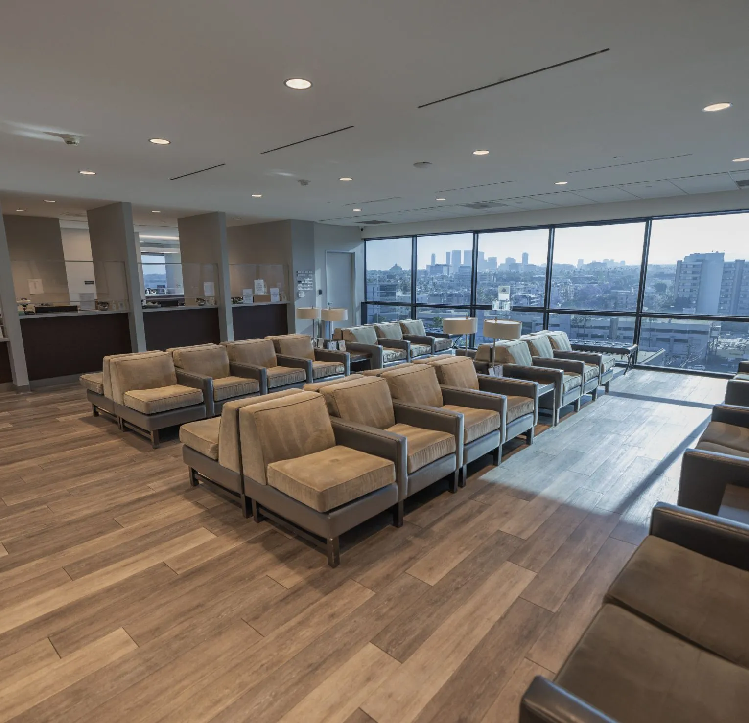
At our convenient Beverly Hills facility, Dr. Parham Yashar offers comprehensive evaluation and treatment for all types of hydrocephalus, including congenital, normal pressure hydrocephalus (NPH), and acquired forms. Using advanced imaging and minimally invasive techniques, Dr. Yashar works to relieve pressure on the brain while preserving cognitive and neurological function.

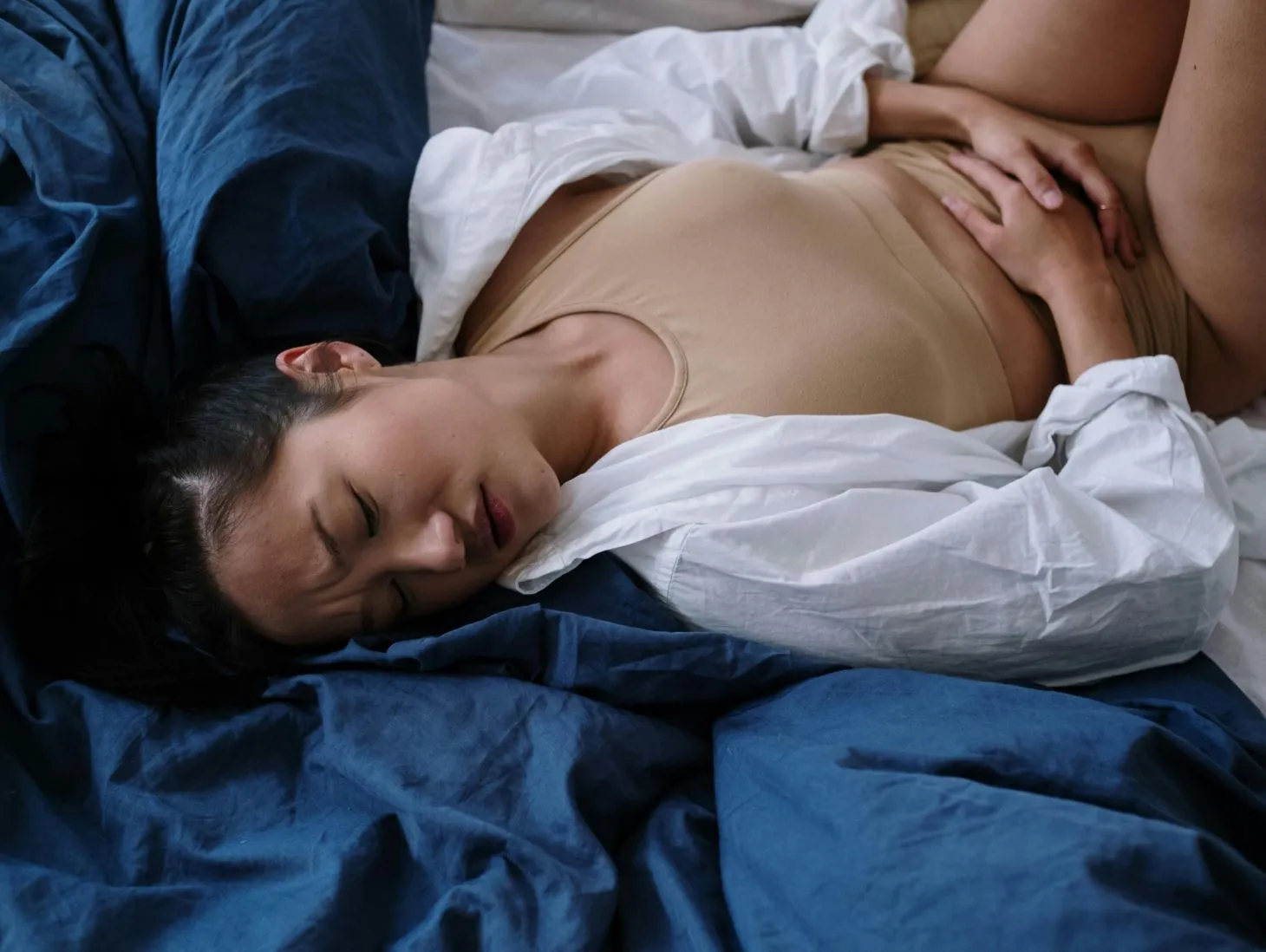
Symptoms of hydrocephalus vary depending on age and severity but may include:
Prompt diagnosis and treatment are key to avoiding long-term complications. Regarded as the best hydrocephalus surgeon in Los Angeles, Dr. Yashar uses high-resolution MRI and CT scans to assess ventricular size and pressure, allowing for accurate diagnosis and individualized care.
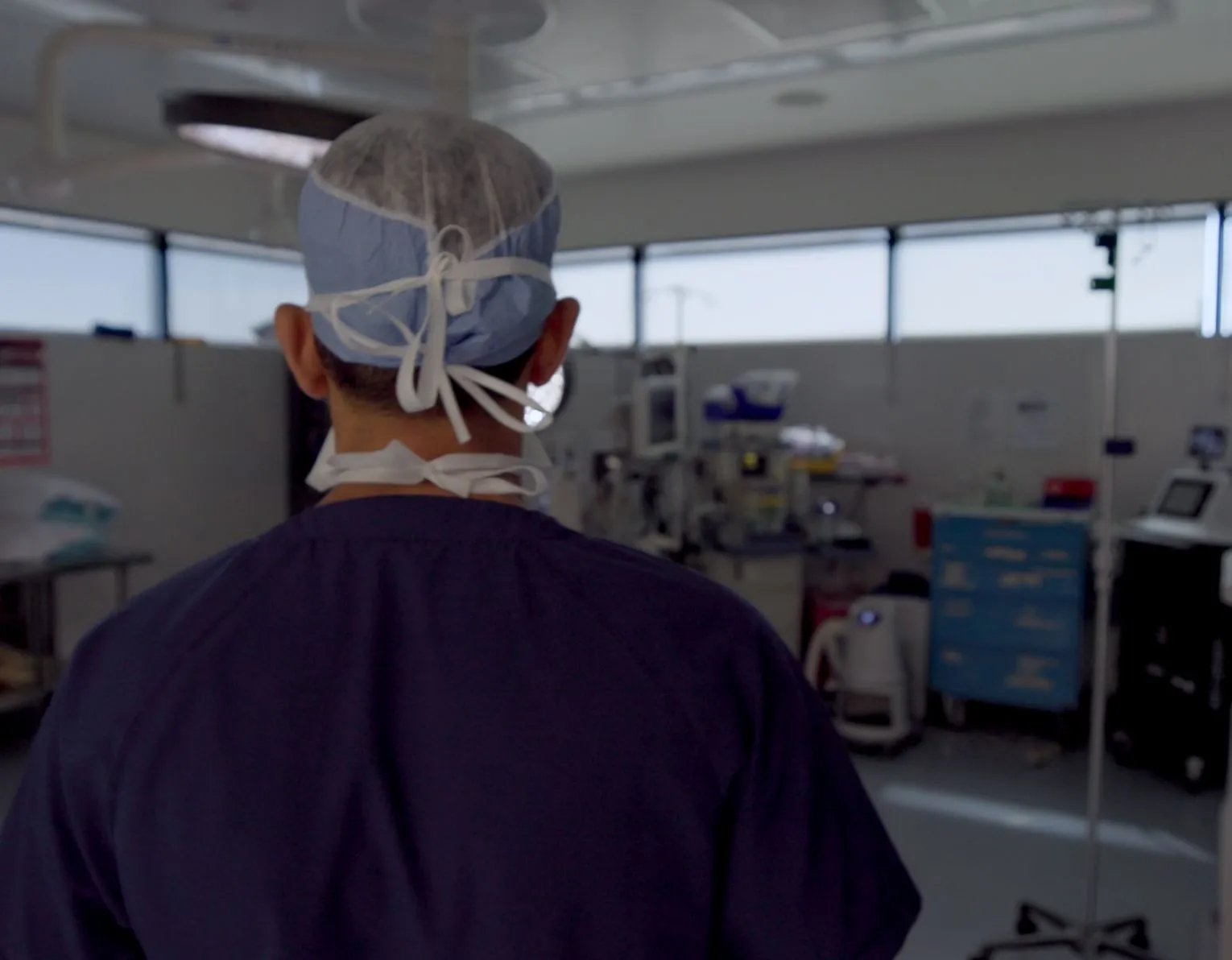
Treatment focuses on diverting or controlling the flow of cerebrospinal fluid to relieve intracranial pressure. Dr. Yashar offers the most effective, minimally invasive surgical options tailored to the patient’s specific type of hydrocephalus and overall health status.
Surgical treatments include:
Dr. Yashar utilizes image-guided technology and microsurgical tools to ensure maximum safety, accuracy, and long-term effectiveness.


While there is no cure, hydrocephalus can often be managed successfully with surgery. Many patients go on to lead normal, independent lives with proper treatment and follow-up.
Modern shunt systems can last for years, but they may require adjustment or replacement over time. Regular monitoring is essential.
NPH is a type of hydrocephalus most common in older adults and is often misdiagnosed as dementia or Parkinson’s disease. It presents with a triad of symptoms: gait disturbance, urinary incontinence, and cognitive decline.

Dr. Parham Yashar is a leading board-certified neurosurgeon with extensive experience treating complex neurological conditions, including all forms of hydrocephalus. His practice combines state-of-the-art technology with a commitment to personalized, patient-first care.
If you’re considering where to find the best brain surgeon for hydrocephalus surgery in Los Angeles, our practice is conveniently located in Beverly Hills, just minutes from Cedars-Sinai, serving patients across Southern California. We provide accurate diagnoses, minimally invasive treatment, and compassionate support through every step of your healing journey.
Take control of your brain’s health today by scheduling a consultation with the best minimally invasive brain surgeon in Los Angeles and get the finest hydrocephalus care you deserve.


Please complete and submit the form below and a member of our staff will contact you shortly.